Although the world is becoming mostly sedentary, our bodies still require a wide variety of daily movements in order to work well. Many of us struggle to get regular exercise, but even that can fall short of nourishing the body from head to toe. How can we move more—a lot more—when we have sore, stiff parts and overly busy lifestyles? Join Katy Bowman M.S., biomechanist, author, and movement educator as she combines big-picture lessons on biomechanics, kinesiology, physiology, and natural hu ...
…
continue reading
เนื้อหาจัดทำโดย The Physician Assistant Life | Smarty PANCE เนื้อหาพอดแคสต์ทั้งหมด รวมถึงตอน กราฟิก และคำอธิบายพอดแคสต์ได้รับการอัปโหลดและจัดเตรียมโดย The Physician Assistant Life | Smarty PANCE หรือพันธมิตรแพลตฟอร์มพอดแคสต์โดยตรง หากคุณเชื่อว่ามีบุคคลอื่นใช้งานที่มีลิขสิทธิ์ของคุณโดยไม่ได้รับอนุญาต คุณสามารถปฏิบัติตามขั้นตอนที่อธิบายไว้ที่นี่ https://th.player.fm/legal
ที่คล้ายกับ The Audio PANCE and PANRE Physician Assistant Board Review Podcast
The Art of Charm is where self-motivated people, just like you, come to learn from the company’s coaches about to how to master human dynamics, relationships, and becoming your best self with the help of Johnny and AJ, the company’s founders. Johnny and AJ bring their 11 years of coaching experience from their famous Bootcamps, where they host clients in Los Angeles from all over the world and they share their stories, best practices and themselves on this weekly podcast. Not only does The A ...
…
continue reading
Ever wondered what makes great go-to-market leaders grow, even when the going gets tough? We have, too. And we’re on a mission to uncover the magic that makes that growth happen. This is Go-to-Market Magic, the show where we talk to go-to-market leaders and visionaries about the “aha!” moments they experience and the pivotal decisions they’ve made, all in the name of growth. And we’re not just talking about revenue growth that goes up and to the right — we’ll also discuss how they improve th ...
…
continue reading
PT Inquest is an online journal club. Hosted by Jason Tuori, Megan Graham, and Chris Juneau, the show looks at an article every week and discusses how it applies to current physical therapy practice.
…
continue reading
Welcome to the Success Story Podcast, hosted by entrepreneur, business executive, author, educator & speaker, Scott D. Clary (@scottdclary). On this podcast, you'll find interviews, Q&A, keynote presentations & conversations on sales, marketing, business, startups and entrepreneurship. Scott will discuss some of the lessons he's learned over his own career, as well as have candid interviews with execs, celebrities, notable figures and politicians. All who have achieved success through both w ...
…
continue reading
Welcome to Almost 30 - a supportive space to fuel your conscious evolution. Join us, LA-based best friends Krista Williams and Lindsey Simcik, for heart-centered, hilarious conversations and real, raw, impactful interviews with brilliant guests. We dive deep into topics like modern spirituality to health and wellness, aliens to entrepreneurship, social justice, and self development. With every episode, our mission is to empower you, expand what you think is possible and, make you laugh - a l ...
…
continue reading
Join expert voices from Barbell Logic and others from the world of strength for resources to help you get strong for life. Get coaching options and more educational content at barbell-logic.com.
…
continue reading
Montgomery & Co. is a weekly podcast where WNBA champion and part-owner of the Atlanta Dream Renee Montgomery is joined by her mother Bertela Montgomery, her sister Nicole Young and her wife Sirena Grace as they chart a unique path through the business world as four black and brown women keeping their family first at all times. Both insightful and compelling, this one-of-a-kind talk show combines interviews with some of the world’s top innovators & entrepreneurs with sports and culture conve ...
…
continue reading
Radio Advisory is your weekly download on how to untangle healthcare's most pressing challenges, powered by 40 years of Advisory Board research. Whether it's workforce shortages, industry disruptors, or health equity strategy, we're here to help. Our hosts and seasoned researchers talk with industry experts to equip you with knowledge to confront today’s unanswered questions in healthcare. New episodes drop every Tuesday. | www.advisory.com
…
continue reading
From the stuff your mother never told you, to the stuff your doctor never learned, On Health features taboo-busting conversations that demystify and de-stigmatize our bodies, all while bridging the gap between conventional medicine and wellness. Join Yale-trained MD & midwife Aviva Romm and her line-up of expert guests as they discuss everything from periods to menopause, sex to reproductive health politics, and motherhood to mental health. Each week, Dr. Romm will be exploring the science a ...
…
continue reading
Player FM - แอป Podcast
ออฟไลน์ด้วยแอป Player FM !
ออฟไลน์ด้วยแอป Player FM !

))
Podcast Episode 75: Ten FREE PANCE and PANRE Audio Board Review Questions
Manage episode 243658459 series 97199
เนื้อหาจัดทำโดย The Physician Assistant Life | Smarty PANCE เนื้อหาพอดแคสต์ทั้งหมด รวมถึงตอน กราฟิก และคำอธิบายพอดแคสต์ได้รับการอัปโหลดและจัดเตรียมโดย The Physician Assistant Life | Smarty PANCE หรือพันธมิตรแพลตฟอร์มพอดแคสต์โดยตรง หากคุณเชื่อว่ามีบุคคลอื่นใช้งานที่มีลิขสิทธิ์ของคุณโดยไม่ได้รับอนุญาต คุณสามารถปฏิบัติตามขั้นตอนที่อธิบายไว้ที่นี่ https://th.player.fm/legal

Welcome to episode 75 of the Audio PANCE and PANRE PA Board Review Podcast.
Join me as I cover ten PANCE and PANRE Board review questions from the SMARTYPANCE course content following the NCCPA content blueprint (download the FREE cheat sheet).
This week we will be covering ten general board review questions based on the NCCPA PANCE and PANRE Content Blueprints.
Below you will find an interactive exam to complement the podcast.
The Audio PANCE and PANRE Physician Assistant Board Review Podcast
I hope you enjoy this free audio component to the examination portion of this site. The full board review includes over 2,000 interactive board review questions and is available to all members of the PANCE and PANRE Academy and Smarty PANCE.
- You can download and listen to past FREE episodes here, on iTunes, on Google Play Music or Stitcher Radio.
- You can listen to the latest episode, take an interactive quiz and download your results below.
Listen Carefully Then Take The Practice Exam
If you can’t see the audio player click here to listen to the full episode.
Podcast Episode 75: Ten Question PANCE and PANRE Podcast Quiz
The following questions are linked to NCCPA Content Blueprint lessons from the Smarty PANCE and PANRE Board Review Website. If you are a member you will be able to log in and view this interactive video lesson.
1. A 5-year-old girl is brought to the emergency department after drinking a bottle of drain cleaner. It is unknown how much the child drank. She has a past medical history of Down syndrome and obesity. The patient’s vitals are unremarkable. Physical exam is notable for a child in no acute distress. She is tolerating her oral secretions and is interactive. Inspection of the oropharynx is unremarkable. Which of the following is the appropriate management of this patient?
- Dilute hydrochloric acid
- Endoscopy
- Intubation
- Observation
- Polyethylene glycol
Click here to see the answer
Answer: B. Endoscopy
Ingestion of caustic fluids (acid or alkali) such as drain cleaner may lead to esophageal damage and stricture. Any patient presenting after caustic ingestion should have an endoscopy performed between 12 and 24 hours of presentation. Injury due to ingestion of alkaline fluids such as drain/oven cleaner occurs rapidly in the first minutes to hours and is characterized by liquefactive necrosis of the esophageal tissue. Subsequently, esophageal strictures form due to scarring of the affected tissue. Patients should immediately be resuscitated following the ABC’s (airway, breathing, and circulation). Subsequent management involves endoscopy (typically within 12 to 24 hours of ingestion) to assess the extent of the damage and plan any further treatment that may be needed.
Incorrect Answers:
- Answer 1: Dilute hydrochloric acid or administration of any agent to titrate the ingestion is always contraindicated as this will lead to more tissue damage.
- Answer 3: Intubation would be indicated if the patient was failing to protect their airway or if they had impending airway loss. This well-appearing patient has no airway concerns.
- Answer 4: Observation is certainly a part of this patient’s care; however, she must also receive endoscopy to assess for any esophageal/GI tract injury.
- Answer 5: Polyethylene glycol or whole bowel irrigation has limited indications. It may be used to pass bags of drugs if ingested by a packer/stuffer but would not aid in the management of caustic ingestion.
Review NCCPA Blueprint Topic: Ingestion of toxic substances or foreign bodies
2. A 27-year-old man presents to the emergency department after a motor vehicle collision. The patient was a front seat unrestrained driver in a head-on collision. The patient’s echocardiogram (ECG) is notable only for sinus tachycardia. His temperature is 99.5°F (37.5°C), blood pressure is 107/58 mmHg, pulse is 120/min, respirations are 17/min, and oxygen saturation is 98% on room air. The patient is given 2 liters of Ringer lactate solution and morphine. Initial workup demonstrates that the patient’s pulmonary capillary wedge pressure and troponins are elevated. The patient is currently complaining of chest pain. Physical exam is notable for an uncomfortable young man with bruising over his chest wall. Which of the following is the most likely diagnosis?
- Cardiac contusion
- Hemorrhage
- Myocardial infarction
- Pulmonary contusion
- Takotsubo cardiomyopathy
Click here to see the answer
Answer: A. Cardiac contusion
This patient is presenting after blunt chest trauma (which is common in motor vehicle accidents) with chest pain, elevated troponins, and an elevated pulmonary capillary wedge pressure suggesting a diagnosis of a cardiac contusion. A cardiac contusion is a blunt injury to the heart which can disrupt the mechanical and electrical function of the heart. There will typically be visible signs of chest trauma such as bruising and the patient will often complain of chest pain and dyspnea. The ECG can be unremarkable, demonstrate sinus tachycardia, or even demonstrate more severe dysrhythmias such as supraventricular tachycardia, atrial fibrillation, and a right bundle branch block. Initial cardiac troponins can be elevated. In the setting of any ECG abnormality or elevated troponins, patients with a suspected diagnosis of a cardiac contusion should be admitted to the hospital and observed until clinically stable with normalization of their troponins.
Incorrect Answers:
- Answer 2: Hemorrhage would present with hypotension, tachycardia, and a decreased pulmonary capillary wedge pressure. Internal bleeding could be assessed with a FAST exam which assesses for pleural sliding, pericardial fluid and cardiac function, fluid in the hepatorenal and splenorenal recesses, and fluid surrounding the bladder.
- Answer 3: Myocardial infarction presents with chest pain, shortness of breath, ST elevation on ECG and elevated cardiac troponins. This patient’s elevated troponins are likely secondary to blunt trauma to the heart as he has no risk factors for ischemic heart disease and is young.
- Answer 4: Pulmonary contusion presents with chest pain, hypoxia, and patchy opacities on chest radiography which may not be initially apparent. Management for a pulmonary contusion is typically supportive in nature and to adequately control the patient’s pain.
- Answer 5: Takotsubo cardiomyopathy presents with chest pain and dyspnea as well as ST elevation on ECG without reciprocal changes. Global hypokinesis of the heart can be seen on echocardiography and minor elevations in troponins can be present. The typical precipitating event for Takotsubo cardiomyopathy is an emotionally stressful life event and the patient is typically a woman in contrast to this male patient who has a mechanism of injury that supports a diagnosis of a cardiac contusion.
Review NCCPA Blueprint Topic: Chest/Rib Fractures and Trauma
3. A 33-year-old woman presents to her primary care PA for a wellness check-up. She states that recently she has been feeling well other than headaches that occur occasionally, which improve with ibuprofen and rest. She has a past medical history of hypertension and headaches and is currently taking hydrochlorothiazide. Her temperature is 99.2°F (37.3°C), blood pressure is 157/108 mmHg, pulse is 90/min, respirations are 14/min, and oxygen saturation is 98% on room air. Physical exam reveals a young woman who appears healthy. A normal S1 and S2 are auscultated on cardiac exam, and her lungs are clear with good air movement bilaterally. From her previous visit, it was determined that she has an elevated aldosterone and low renin level. Laboratory values are ordered as seen below.
Serum:
Na+: 139 mEq/L
Cl-: 100 mEq/L
K+: 3.7 mEq/L
HCO3-: 29 mEq/L
BUN: 20 mg/dL
Creatinine: 1.1 mg/dL
Which of the following is the most likely diagnosis?
- Benign essential hypertension
- Cushing syndrome
- Narrowing of the renal arteries
- Pheochromocytoma
- Primary hyperaldosteronism
Click here to see the answer
Answer: E. Primary hyperaldosteronism
This patient is presenting with hypertension refractory to a diuretic with a decreased potassium, a decreased renin level, and an increased aldosterone level suggesting a diagnosis of primary hyperaldosteronism.
Primary hyperaldosteronism occurs when the adrenal gland produces excess aldosterone. Aldosterone has the effect in the kidney of absorbing sodium (and thus water) and wasting potassium and hydrogen. Thus, hyperaldosteronism can lead to hypertension, hypokalemia, and a metabolic alkalosis. In primary hyperaldosteronism, the high blood pressure is detected by the kidney, and thus renin levels are decreased in the setting of an elevated aldosterone.
Incorrect Answers:
- Answer 1: Benign essential hypertension typically occurs in overweight patients and has no clear underlying cause. It would not be associated with elevated aldosterone levels.
- Answer 2: Cushing syndrome would be associated with weight gain, limb muscle atrophy, and striae on dermatological exam in the setting of a decreased renin and aldosterone.
- Answer 3: Narrowing of the renal arteries (renal artery stenosis) would present with refractory hypertension in the setting of an elevated renin and aldosterone level.
- Answer 4: Pheochromocytoma would present with severe episodic hypertension and headaches secondary to an adrenal mass that releases catecholamines.
Summary: Primary hyperaldosteronism presents with hypertension, a decreased renin and potassium level, a metabolic alkalosis, and an increased aldosterone level.
Review NCCPA Blueprint Topic: Secondary hypertension
4. A 24-year-old man is brought in to the emergency room after being retrieved by firefighters from a burning building. The patient is responding coherently to questions but reports pain secondary to a burn on his leg. He states he also has a headache and feels dizzy. His temperature is 98.5°F (36.9°C), blood pressure is 129/66 mmHg, pulse is 126/min, respirations are 14/min, and oxygen saturation is 99% on room air. Physical exam is notable for a confused young man with dry and flushed skin. The cardiopulmonary exam reveals a normal S1 and S2, as well as clear breath sounds bilaterally. The patient’s neurological exam is within normal limits. Towards the end of his exam, the patient begins vomiting. The dermatologic exam reveals a superficial burn covering 1% of the patient’s body over his right leg. Which of the following is the best next step in management for this patient?
- 100% oxygen
- CT scan of the head
- Hydroxocobalamin
- Normal saline
- Ondansetron
Click here to see the answer
Answer: A. 100% oxygen
This patient is presenting after being rescued from a fire with confusion, headache, nausea, vomiting, and flushed skin suggesting a diagnosis of carbon monoxide poisoning which should be treated with 100% oxygen.
Carbon monoxide exposure is common in fires and in patients who heat their house with an old-fashioned wood stove. Carbon monoxide binds to hemoglobin with a higher affinity than oxygen thus displacing it leading to symptoms. Carbon monoxide poisoning presents with confusion, headache, altered mental status, nausea, vomiting, and cherry-red skin. Pulse oximetry is often normal in these patients as the device detects hemoglobin bound to oxygen or carbon monoxide similarly. A carboxyhemoglobin level can be obtained to confirm the diagnosis in these patients; however, patients presenting with a clinical picture supportive of carbon monoxide poisoning should be treated with 100% (or hyperbaric) oxygen. Sequelae of carbon monoxide poisoning should be treated supportively, including dantrolene for increased muscle activity or benzodiazepines for seizure activity.
Incorrect Answers:
- Answer 2: CT scan of the head would be indicated if a patient presented with head trauma followed by altered mental status, nausea, and vomiting. This patient’s neurological abnormalities, as well as his nausea and vomiting, can be explained by his carbon monoxide poisoning.
- Answer 3: Hydroxocobalamin is the treatment of choice for cyanide poisoning which is also associated with exposure to fires. Patients will present with weakness, malaise, headache, dizziness, nausea, vomiting, and shortness of breath. Though this diagnosis is possible in this patient, a more likely diagnosis both epidemiologically and given his flushed skin is carbon monoxide poisoning. This patient will need empiric treatment for cyanide poisoning, however, a more dire intervention for the most likely diagnosis is putting the patient on oxygen to simultaneously and rapidly improve his respiratory status and treat his carbon monoxide poisoning.
- Answer 4: Normal saline may be necessary to stabilize this patient’s blood pressure; however, his tachycardia is likely secondary to his pain and a more important initial intervention is stabilizing the patient’s respiratory status including administering oxygen for this patient’s carbon monoxide poisoning.
- Answer 5: Ondansetron only treats this patient’s symptoms of nausea but does not address their respiratory status which should be treated promptly and prior to treating other concerns.
Summary: The treatment of carbon monoxide poisoning is 100% oxygen.
Review NCCPA Blueprint Topic: Burns (ReelDx)
5. A 42-year-old woman is brought to the emergency department after a motor vehicle accident. She complains of lower back pain and some minor abdominal pain. The patient has a past medical history of obesity and type II diabetes. Her current medications include atorvastatin, metformin, and glyburide. A FAST exam is performed in the trauma bay and does not reveal any signs of intra-abdominal bleeding. Her temperature is 98.2°F (36.8°C), blood pressure is 130/77 mmHg, pulse is 90/min, respirations are 16/min, and oxygen saturation is 99% on room air. Ultrasound findings are notable for multiple gallstones in the gallbladder. The patient is given naproxen. Which of the following is the best next step in management?
- CT scan of the abdomen
- NPO, IV fluids, and broad-spectrum antibiotics
- Perform a cholecystectomy this hospital visit
- Schedule an outpatient cholecystectomy
- Supportive therapy
Click here to see the answer
Answer: E. Supportive therapy
This patient is presenting with asymptomatic gallstones discovered incidentally. Asymptomatic gallstones do not need to be managed with a cholecystectomy.
Acute cholecystitis classically presents with right upper quadrant abdominal pain that presents in a fat, fertile, female in her forties. Once the diagnosis is confirmed with ultrasound, “cooling off” of the gallbladder is necessary (keeping the patient NPO and IV fluids) followed by a cholecystectomy that hospital visit. However, if asymptomatic gallstones are discovered incidentally, there is no indication for cholecystectomy. These patients should be managed conservatively.
Incorrect Answers:
- Answer 1: CT scan of the abdomen is unnecessary as there is no indication to perform a CT scan, in particular when the only finding in this assessment was asymptomatic gallstones on ultrasound.
- Answer 2: NPO, IV fluids, and broad spectrum antibiotics is the best initial management of ascending cholangitis. Patients with ascending cholangitis are acutely ill and present with a fever, jaundice, and right upper quadrant tenderness. They require immediate treatment as well as decompression (from interventional radiology), followed by ERCP/cholecystectomy.
- Answer 3: Performing a cholecystectomy this hospital visit is the appropriate management of acute cholecystitis. Acute cholecystitis would present with right upper quadrant abdominal pain in an overweight woman after eating a fatty meal. It is not indicated in the management of asymptomatic gallstones.
- Answer 4: Scheduling an outpatient cholecystectomy is not necessary as this patient is asymptomatic. Outpatient cholecystectomies are not typically performed in the management of acute cholecystitis as most patients do not comply with outpatient recommendations (avoiding triggers like fatty foods and alcohol) and end up returning to the hospital for another flare before their procedure. In certain compliant patients, it may be a viable option.
Review NCCPA Blueprint Topic: Cholelithiasis (ReelDx + Lecture)
6. A 60-year-old woman presents to the emergency department with dizziness. She states it started this morning when she woke up from bed and was severe causing her to vomit. The episode resolved in 1 minute. The patient has a past medical history of hypertension, diabetes, obesity, and atrial fibrillation treated with warfarin and metoprolol. She recently recovered from a cold a few days ago. Her temperature is 99.0°F (37.2°C), blood pressure is 174/99 mmHg, pulse is 115/min, respirations are 12/min, and oxygen saturation is 98% on room air. Physical exam is notable for a well-appearing woman. Her neurological exam including cranial nerves and gait is within normal limits. The patient is laid flat in the bed which causes an episode of dizziness with notable nystagmus and vomiting. She feels better after 1 minute. The patient’s ECG is within normal limits. Lab values are notable for an INR of 3.5. Which of the following is the most likely etiology of this patient’s symptoms?
- Canalithiasis
- Increased endolymph production
- Inflammation of the vestibular apparatus
- Inflammation of the vestibulocochlear apparatus
- Vertebrobasilar insufficiency
Click here to see the answer
Answer: A. Canalithiasis
This patient is presenting with intermittent, severe vertigo which is provoked by position changes which is most consistent with benign paroxysmal positional vertigo (BPPV). BPPV is commonly caused by canalithiasis.
Benign paroxysmal positional vertigo (BPPV) is a common form of peripheral vertigo that results from a dislodged piece of otolith (called otoconia when dislodged) causing disturbances in the semicircular canals. The presentation of BPPV involves sudden and episodic vertigo with head movements that lasts for seconds to minutes accompanied by nausea and vomiting. Physical exam will demonstrate a horizontal nystagmus with specific head postures (such as the Dix-Hallpike maneuver). Treatment involves repositioning exercises (such as the Epley maneuver) as well as meclizine or diphenhydramine for symptomatic control.
Incorrect Answers:
- Answer 2: Increased endolymph production describes Meniere disease which presents with chronic symptoms including hearing loss and ear fullness and intermittent episodes of vertigo. Treatment involves diuretics and salt restriction.
- Answers 3-4: Inflammation of the vestibular apparatus and the vestibulocochlear apparatus describes vestibular neuritis (sustained/persistent vertigo after a cold) and labyrinthitis (sustained/persistent vertigo and hearing loss after a cold), respectively. This condition will resolve on its own; however, symptoms can be treated with meclizine or diphenhydramine.
- Answer 5: Vertebrobasilar insufficiency can present with syncope or if there is a hemorrhage/ischemia, may present in an elderly patient with multiple risk factors with sustained and severe vertigo that is sudden onset and associated with dysarthria and dystonia.
Summary: Benign paroxysmal positional vertigo is commonly caused by canalithiasis.
Review NCCPA Blueprint Topic: Vertigo (ReelDx + Lecture)
7. An 18-year-old male presents to his primary care provider with his parents for a sports physical. He was last seen in the clinic several months ago when he was diagnosed with attention deficit hyperactivity disorder (ADHD). He was started on methylphenidate at that time, and the patient now reports improvement in his ability to concentrate in school and at home. He hopes to play baseball in college and has begun lifting weights daily in preparation for baseball season. The patient reports that he eats a healthy diet to fuel his exercise regimen. His parents have no concerns and are pleased with the recent improvement in his grades. On physical exam, the patient has tall stature with average muscle mass for his age. He has no dysmorphic features. His chest has a normal appearance other than mild gynecomastia. The patient has sparse facial hair and a moderate amount of coarse pubic hair that extends across the pubis and spares the medial thighs. His testes are small and firm. Due to the latter, laboratory testing is performed and reveals the following:
- Follicle-stimulating hormone (FSH): 42 mIU/mL (Reference range: 4-25 mIU/mL)
- Luteinizing hormone (LH): 38 mIU/mL (Reference range: 6-23 mIU/mL)
Which of the following is the most likely etiology of this patient’s presentation?
- Anabolic steroid use
- CGG trinucleotide repeat disorder
- CTG trinucleotide repeat disorder
- Failure of neuronal migration
- Meiotic nondisjunction
Click here to see the answer
Answer: E. Meiotic nondisjunction
This patient presents with tall stature, gynecomastia, and small testes with elevated FSH and LH, which suggests a diagnosis of Klinefelter syndrome. Klinefelter syndrome is usually caused by meiotic nondisjunction that results in a 47,XXY genotype.
Klinefelter syndrome is the most common cause of primary hypogonadism. Patients with Klinefelter syndrome present with tall stature, neurocognitive difficulties (ADHD) and features of hypogonadism including gynecomastia, small testes, small phallus, hypospadias, underdeveloped secondary sex characteristics, and cryptorchidism. Patients without hypospadias or cryptorchidism are often not diagnosed until after puberty, when the symptoms of gynecomastia and small testes become more prominent. Because the hypogonadism in Klinefelter syndrome is caused by testicular fibrosis, laboratory results demonstrate a low testosterone and elevated FSH and LH.
Incorrect Answers:
- Answer 1: Anabolic steroid use causes decreased levels of FSH and LH due to the suppression of GnRH release by the hypothalamus, which in turn suppresses FSH and LH release by the pituitary gland. Anabolic steroid use would not present with signs of hypogonadism.
- Answer 2: The CGG trinucleotide repeat disorder characterizes Fragile X syndrome. Fragile X presents with macroorchidism rather than hypogonadism, and patients typically have dysmorphic features of a long, narrow face with large ears, prominent forehead, and prominent chin. Fragile X is the most common cause of inherited intellectual disability.
- Answer 3: The CTG trinucleotide repeat disorder characterizes myotonic dystrophy. Although myotonic dystrophy presents with hypogonadism, patients would also present with symptoms of progressive weakness, such as facial weakness, dysphagia, or hand grip weakness.
- Answer 4: Failure of neuronal migration characterizes Kallmann syndrome. Kallmann syndrome presents with the classic symptoms of loss of smell and hypogonadism, but patients with Kallmann syndrome have a low FSH and LH.
Summary: Klinefelter syndrome results in primary hypogonadism and presents with tall stature, gynecomastia, small testes, a small phallus, hypospadias, and cryptorchidism.
Review NCCPA Blueprint Topic: Hypogonadism
8. A 65-year-old man presents to the emergency department for sudden weakness. The patient states that he was at home enjoying his morning coffee when his symptoms began. He says that his left arm suddenly felt very odd and weak thus prompting him to come to the ED. The patient has a past medical history of diabetes, COPD, hypertension, anxiety, alcohol abuse, and PTSD. He recently fell off a horse while horseback riding but claims to not have experienced any significant injuries. He typically drinks 5-7 drinks per day and his last drink was yesterday afternoon. His current medications include insulin, metformin, atorvastatin, lisinopril, albuterol, and fluoxetine. His temperature is 99.5°F (37.5°C), blood pressure is 177/118 mmHg, pulse is 120/min, respirations are 18/min, and oxygen saturation is 93% on room air. On physical exam, you note an elderly man who is mildly confused. The cardiopulmonary exam demonstrates bilateral expiratory wheezes and a systolic murmur along the right upper sternal border that radiates to the carotids. Neurological exam reveals cranial nerves II-XII as grossly intact with finger-nose exam mildly abnormal on the left and heel-shin exam within normal limits. The patient has 5/5 strength in his right arm and 3/5 strength in his left arm. The patient struggles to manipulate objects such as a pen with his left hand. The patient is given a dose of diazepam and started on IV fluids. Which of the following is the most likely diagnosis in this patient?
- Berry aneurysm rupture
- Bridging vein tear
- Cerebellar bleeding
- Hypertensive encephalopathy
- Lacunar stroke
Click here to see the answer
Answer: E. Lacunar stroke
This patient is presenting with risk factors and symptoms suggestive of a diagnosis of a lacunar stroke.
Lacunar strokes typically occur in patients with risk factors such as hypertension, diabetes, old age, and smoking. The basal ganglia, pons, and subcortical white matter are commonly affected. The pathophysiology occurs secondary to a small penetrating artery occlusion from hypertensive arteriolar sclerosis, lipohyalinosis, or microatheroma formation. Patients present with neurological deficits that can include pure motor hemiparesis, pure sensory stroke, ataxic hemiparesis, or dysarthria-clumsy hand syndrome.
Incorrect Answers:
- Answer 1: Berry aneurysm rupture describes a subarachnoid hemorrhage that would present with a sudden onset, severe headache.
- Answer 2: Bridging vein tear describes a subdural hematoma which could present with gradual neurological deficits and altered cognition in the setting of recent trauma in an elderly patient/alcoholic.
- Answer 3: Cerebellar bleeding would present with ataxic gait and an abnormal finger-nose and heel-shin exam.
- Answer 4: Hypertensive encephalopathy presents with general CNS dysfunction which can include headache, irritability, nausea/vomiting, disturbances of consciousness, and seizures.
Review NCCPA Blueprint Topic: Stroke (ReelDx + Lecture)
9. A 26-year-old woman presents to the emergency department with abdominal pain. She states that she was walking up the stairs at work when she felt sudden and severe abdominal pain followed by nausea and vomiting. Her past medical history is noncontributory and she is not currently taking any medications. Her temperature is 99.7°F (37.6°C), blood pressure is 122/78 mmHg, pulse is 120/min, respirations are 17/min, and oxygen saturation is 98% on room air. Physical exam is notable for an absence of abdominal tenderness, a left adnexal mass, and left adnexal tenderness. A transvaginal ultrasound demonstrates free fluid surrounding the ovary with edema and the presence of doppler flow. A urinary pregnancy test is negative. The patient’s symptoms persisted after ibuprofen and acetaminophen. Which of the following is the best next step in management?
- CT scan of the abdomen
- Laparoscopy
- Laparotomy
- MRI of the pelvis
- Observation and serial abdominal exams
Click here to see the answer
Answer: B. Laparoscopy
This patient is presenting with sudden onset abdominal pain, nausea, vomiting, and free fluid around the ovary with normal blood flow which is still concerning for ovarian torsion that should be managed with laparoscopy. Ovarian torsion occurs when the ovary twists around its blood supply causing ischemia and necrosis. Patients are typically young women who experience sudden/severe abdominal or vaginal pain, blood per vagina, a left adnexal mass with adnexal tenderness, and a negative urine pregnancy test. The best initial test is a transvaginal ultrasound with Doppler which can show blood flow to the ovary as well as ovarian enlargement and edema (signs of ischemia). Even in the setting of normal blood flow, torsion is possible as the ovary may twist and untwist. Patients presenting with symptoms concerning for ovarian torsion should be managed with laparoscopy to definitively treat and salvage the ovary.
Incorrect Answers:
- Answer 1: CT scan of the abdomen would not be necessary as this patient has no abdominal tenderness and has physical exam and ultrasound findings concerning for ovarian torsion which should be managed surgically.
- Answer 3: Laparotomy is an open procedure that would be indicated if the patient suddenly decompensated and needed more aggressive management to stop acute bleeding.
- Answer 4: MRI of the pelvis would be a very accurate test for diagnosing pelvic pathology; however, it is not necessary in this patient who has a history and findings concerning for ovarian torsion. Performing an MRI would only delay treatment.
- Answer 5: Observation and serial abdominal exams would be the appropriate management of a patient with abdominal pain with a clear etiology after a CT scan had ruled out any serious pathology. This patient’s likely ovarian torsion should not be observed.
Summary: Ovarian torsion should be treated with laparoscopy.
Review NCCPA Blueprint Topic: Ovarian torsion
10. A 72-year-old man is brought into the emergency department by emergency medical services. He looks disheveled and states that he is homeless. He has bruising over his arms and legs and states that he does not have a regular source of nutrition. He denies prior medical conditions but states that he still smokes one pack of cigarettes per day. On exam, the patient’s vital signs are normal, but he appears extremely malnourished. His gums are swollen and bleeding and his tongue is unusually smooth. The hair on his arms is pinwheel-shaped. What is the most likely cause?
- Iron deficiency
- Vitamin B3 deficiency
- Vitamin C deficiency
- Vitamin B12 deficiency
- Vitamin K deficiency
Click here to see the answer
Answer: C. Vitamin C deficiency
An elderly, malnourished, cigarette-smoking male with swollen gums, bruising, and corkscrew hair is most likely suffering from vitamin C deficiency.
Though vitamin C deficiency, or scurvy, is uncommon in the developed world, cases in developing countries still exist. Typically, these cases occur in those who are very old or very young due to the inability to feed themselves properly. Since vitamin C, or ascorbic acid, is found in citrus fruits and green vegetables, deficiency in these foods or consumption of these foods with denatured vitamins (due to over-boiling) can result in deficiency. Those who smoke cigarettes have also been found to be more deficient in vitamin C.
Incorrect Answers:
- Answer 1: Iron deficiency anemia would most commonly be asymptomatic, and if serious, would present with symptoms of fatigue and shortness of breath. It would not present with glossitis or corkscrew hair.
- Answer 2: Vitamin B3, or niacin, can present with glossitis, but would typically also have the “3 D’s”: diarrhea, dermatitis, and dementia.
- Answer 4: Vitamin B12 deficiency can also present with glossitis, but it would also have physical exam signs such as numbness and parasthesias of the extremities along with ataxia.
- Answer 5: Vitamin K deficiency can present with bleeding and bruising, but it would not present with the other symptoms typically associated with scurvy.
Review NCCPA Blueprint Topic: Hypervitaminosis/hypovitaminosis
[spoiler title=”CLooking for all the podcast episodes?
This FREE series is limited to every other episode, you can download and enjoy the complete audio series by joining The PANCE and PANRE Exam Academy + SMARTYPANCE
I will be releasing new episodes every few weeks. The Academy is discounted, so sign up now.
Resources and Links From The Show
- My list of recommended PANCE and PANRE review books
- Interactive PANCE and PANRE NCCPA Exam Content Blueprints
- Sign up for the FREE Daily PANCE and PANRE email series
- Join the Smarty PANCE NCCPA Content Blueprint Website + The PA Life Academy
- Get 20% of any Picmonic membership by using this link
- Use Code “PALIFE” and get 10% OFF THE RUTGERS PANCE AND PANRE REVIEW COURSE
This Podcast is also available on iTunes and Stitcher Radio for Android
- iTunes: The Audio PANCE and PANRE Podcast iTunes
- Stitcher Radio: The Audio PANCE and PANRE Podcast Stitcher
- Google Play: The Audio PANCE and PANRE Podcast Google Play
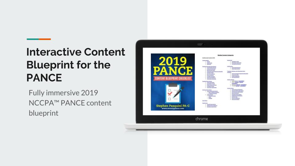
Download The Content Blueprint Checklist
Follow this link to download your FREE copy of the Content Blueprint Checklist
Print it up and start crossing out the topics you understand, marking the ones you don’t and making notes of key terms you should remember. The PDF version is interactive and linked directly to the individual lessons on SMARTY PANCE.
Download for PANCE Download for PANRE
The post Podcast Episode 75: Ten FREE PANCE and PANRE Audio Board Review Questions appeared first on The Audio PANCE and PANRE.
67 ตอน
Podcast Episode 75: Ten FREE PANCE and PANRE Audio Board Review Questions
The Audio PANCE and PANRE Physician Assistant Board Review Podcast
Manage episode 243658459 series 97199
เนื้อหาจัดทำโดย The Physician Assistant Life | Smarty PANCE เนื้อหาพอดแคสต์ทั้งหมด รวมถึงตอน กราฟิก และคำอธิบายพอดแคสต์ได้รับการอัปโหลดและจัดเตรียมโดย The Physician Assistant Life | Smarty PANCE หรือพันธมิตรแพลตฟอร์มพอดแคสต์โดยตรง หากคุณเชื่อว่ามีบุคคลอื่นใช้งานที่มีลิขสิทธิ์ของคุณโดยไม่ได้รับอนุญาต คุณสามารถปฏิบัติตามขั้นตอนที่อธิบายไว้ที่นี่ https://th.player.fm/legal
Welcome to episode 75 of the Audio PANCE and PANRE PA Board Review Podcast.
Join me as I cover ten PANCE and PANRE Board review questions from the SMARTYPANCE course content following the NCCPA content blueprint (download the FREE cheat sheet).
This week we will be covering ten general board review questions based on the NCCPA PANCE and PANRE Content Blueprints.
Below you will find an interactive exam to complement the podcast.
The Audio PANCE and PANRE Physician Assistant Board Review Podcast
I hope you enjoy this free audio component to the examination portion of this site. The full board review includes over 2,000 interactive board review questions and is available to all members of the PANCE and PANRE Academy and Smarty PANCE.
- You can download and listen to past FREE episodes here, on iTunes, on Google Play Music or Stitcher Radio.
- You can listen to the latest episode, take an interactive quiz and download your results below.
Listen Carefully Then Take The Practice Exam
If you can’t see the audio player click here to listen to the full episode.
Podcast Episode 75: Ten Question PANCE and PANRE Podcast Quiz
The following questions are linked to NCCPA Content Blueprint lessons from the Smarty PANCE and PANRE Board Review Website. If you are a member you will be able to log in and view this interactive video lesson.
1. A 5-year-old girl is brought to the emergency department after drinking a bottle of drain cleaner. It is unknown how much the child drank. She has a past medical history of Down syndrome and obesity. The patient’s vitals are unremarkable. Physical exam is notable for a child in no acute distress. She is tolerating her oral secretions and is interactive. Inspection of the oropharynx is unremarkable. Which of the following is the appropriate management of this patient?
- Dilute hydrochloric acid
- Endoscopy
- Intubation
- Observation
- Polyethylene glycol
Click here to see the answer
Answer: B. Endoscopy
Ingestion of caustic fluids (acid or alkali) such as drain cleaner may lead to esophageal damage and stricture. Any patient presenting after caustic ingestion should have an endoscopy performed between 12 and 24 hours of presentation. Injury due to ingestion of alkaline fluids such as drain/oven cleaner occurs rapidly in the first minutes to hours and is characterized by liquefactive necrosis of the esophageal tissue. Subsequently, esophageal strictures form due to scarring of the affected tissue. Patients should immediately be resuscitated following the ABC’s (airway, breathing, and circulation). Subsequent management involves endoscopy (typically within 12 to 24 hours of ingestion) to assess the extent of the damage and plan any further treatment that may be needed.
Incorrect Answers:
- Answer 1: Dilute hydrochloric acid or administration of any agent to titrate the ingestion is always contraindicated as this will lead to more tissue damage.
- Answer 3: Intubation would be indicated if the patient was failing to protect their airway or if they had impending airway loss. This well-appearing patient has no airway concerns.
- Answer 4: Observation is certainly a part of this patient’s care; however, she must also receive endoscopy to assess for any esophageal/GI tract injury.
- Answer 5: Polyethylene glycol or whole bowel irrigation has limited indications. It may be used to pass bags of drugs if ingested by a packer/stuffer but would not aid in the management of caustic ingestion.
Review NCCPA Blueprint Topic: Ingestion of toxic substances or foreign bodies
2. A 27-year-old man presents to the emergency department after a motor vehicle collision. The patient was a front seat unrestrained driver in a head-on collision. The patient’s echocardiogram (ECG) is notable only for sinus tachycardia. His temperature is 99.5°F (37.5°C), blood pressure is 107/58 mmHg, pulse is 120/min, respirations are 17/min, and oxygen saturation is 98% on room air. The patient is given 2 liters of Ringer lactate solution and morphine. Initial workup demonstrates that the patient’s pulmonary capillary wedge pressure and troponins are elevated. The patient is currently complaining of chest pain. Physical exam is notable for an uncomfortable young man with bruising over his chest wall. Which of the following is the most likely diagnosis?
- Cardiac contusion
- Hemorrhage
- Myocardial infarction
- Pulmonary contusion
- Takotsubo cardiomyopathy
Click here to see the answer
Answer: A. Cardiac contusion
This patient is presenting after blunt chest trauma (which is common in motor vehicle accidents) with chest pain, elevated troponins, and an elevated pulmonary capillary wedge pressure suggesting a diagnosis of a cardiac contusion. A cardiac contusion is a blunt injury to the heart which can disrupt the mechanical and electrical function of the heart. There will typically be visible signs of chest trauma such as bruising and the patient will often complain of chest pain and dyspnea. The ECG can be unremarkable, demonstrate sinus tachycardia, or even demonstrate more severe dysrhythmias such as supraventricular tachycardia, atrial fibrillation, and a right bundle branch block. Initial cardiac troponins can be elevated. In the setting of any ECG abnormality or elevated troponins, patients with a suspected diagnosis of a cardiac contusion should be admitted to the hospital and observed until clinically stable with normalization of their troponins.
Incorrect Answers:
- Answer 2: Hemorrhage would present with hypotension, tachycardia, and a decreased pulmonary capillary wedge pressure. Internal bleeding could be assessed with a FAST exam which assesses for pleural sliding, pericardial fluid and cardiac function, fluid in the hepatorenal and splenorenal recesses, and fluid surrounding the bladder.
- Answer 3: Myocardial infarction presents with chest pain, shortness of breath, ST elevation on ECG and elevated cardiac troponins. This patient’s elevated troponins are likely secondary to blunt trauma to the heart as he has no risk factors for ischemic heart disease and is young.
- Answer 4: Pulmonary contusion presents with chest pain, hypoxia, and patchy opacities on chest radiography which may not be initially apparent. Management for a pulmonary contusion is typically supportive in nature and to adequately control the patient’s pain.
- Answer 5: Takotsubo cardiomyopathy presents with chest pain and dyspnea as well as ST elevation on ECG without reciprocal changes. Global hypokinesis of the heart can be seen on echocardiography and minor elevations in troponins can be present. The typical precipitating event for Takotsubo cardiomyopathy is an emotionally stressful life event and the patient is typically a woman in contrast to this male patient who has a mechanism of injury that supports a diagnosis of a cardiac contusion.
Review NCCPA Blueprint Topic: Chest/Rib Fractures and Trauma
3. A 33-year-old woman presents to her primary care PA for a wellness check-up. She states that recently she has been feeling well other than headaches that occur occasionally, which improve with ibuprofen and rest. She has a past medical history of hypertension and headaches and is currently taking hydrochlorothiazide. Her temperature is 99.2°F (37.3°C), blood pressure is 157/108 mmHg, pulse is 90/min, respirations are 14/min, and oxygen saturation is 98% on room air. Physical exam reveals a young woman who appears healthy. A normal S1 and S2 are auscultated on cardiac exam, and her lungs are clear with good air movement bilaterally. From her previous visit, it was determined that she has an elevated aldosterone and low renin level. Laboratory values are ordered as seen below.
Serum:
Na+: 139 mEq/L
Cl-: 100 mEq/L
K+: 3.7 mEq/L
HCO3-: 29 mEq/L
BUN: 20 mg/dL
Creatinine: 1.1 mg/dL
Which of the following is the most likely diagnosis?
- Benign essential hypertension
- Cushing syndrome
- Narrowing of the renal arteries
- Pheochromocytoma
- Primary hyperaldosteronism
Click here to see the answer
Answer: E. Primary hyperaldosteronism
This patient is presenting with hypertension refractory to a diuretic with a decreased potassium, a decreased renin level, and an increased aldosterone level suggesting a diagnosis of primary hyperaldosteronism.
Primary hyperaldosteronism occurs when the adrenal gland produces excess aldosterone. Aldosterone has the effect in the kidney of absorbing sodium (and thus water) and wasting potassium and hydrogen. Thus, hyperaldosteronism can lead to hypertension, hypokalemia, and a metabolic alkalosis. In primary hyperaldosteronism, the high blood pressure is detected by the kidney, and thus renin levels are decreased in the setting of an elevated aldosterone.
Incorrect Answers:
- Answer 1: Benign essential hypertension typically occurs in overweight patients and has no clear underlying cause. It would not be associated with elevated aldosterone levels.
- Answer 2: Cushing syndrome would be associated with weight gain, limb muscle atrophy, and striae on dermatological exam in the setting of a decreased renin and aldosterone.
- Answer 3: Narrowing of the renal arteries (renal artery stenosis) would present with refractory hypertension in the setting of an elevated renin and aldosterone level.
- Answer 4: Pheochromocytoma would present with severe episodic hypertension and headaches secondary to an adrenal mass that releases catecholamines.
Summary: Primary hyperaldosteronism presents with hypertension, a decreased renin and potassium level, a metabolic alkalosis, and an increased aldosterone level.
Review NCCPA Blueprint Topic: Secondary hypertension
4. A 24-year-old man is brought in to the emergency room after being retrieved by firefighters from a burning building. The patient is responding coherently to questions but reports pain secondary to a burn on his leg. He states he also has a headache and feels dizzy. His temperature is 98.5°F (36.9°C), blood pressure is 129/66 mmHg, pulse is 126/min, respirations are 14/min, and oxygen saturation is 99% on room air. Physical exam is notable for a confused young man with dry and flushed skin. The cardiopulmonary exam reveals a normal S1 and S2, as well as clear breath sounds bilaterally. The patient’s neurological exam is within normal limits. Towards the end of his exam, the patient begins vomiting. The dermatologic exam reveals a superficial burn covering 1% of the patient’s body over his right leg. Which of the following is the best next step in management for this patient?
- 100% oxygen
- CT scan of the head
- Hydroxocobalamin
- Normal saline
- Ondansetron
Click here to see the answer
Answer: A. 100% oxygen
This patient is presenting after being rescued from a fire with confusion, headache, nausea, vomiting, and flushed skin suggesting a diagnosis of carbon monoxide poisoning which should be treated with 100% oxygen.
Carbon monoxide exposure is common in fires and in patients who heat their house with an old-fashioned wood stove. Carbon monoxide binds to hemoglobin with a higher affinity than oxygen thus displacing it leading to symptoms. Carbon monoxide poisoning presents with confusion, headache, altered mental status, nausea, vomiting, and cherry-red skin. Pulse oximetry is often normal in these patients as the device detects hemoglobin bound to oxygen or carbon monoxide similarly. A carboxyhemoglobin level can be obtained to confirm the diagnosis in these patients; however, patients presenting with a clinical picture supportive of carbon monoxide poisoning should be treated with 100% (or hyperbaric) oxygen. Sequelae of carbon monoxide poisoning should be treated supportively, including dantrolene for increased muscle activity or benzodiazepines for seizure activity.
Incorrect Answers:
- Answer 2: CT scan of the head would be indicated if a patient presented with head trauma followed by altered mental status, nausea, and vomiting. This patient’s neurological abnormalities, as well as his nausea and vomiting, can be explained by his carbon monoxide poisoning.
- Answer 3: Hydroxocobalamin is the treatment of choice for cyanide poisoning which is also associated with exposure to fires. Patients will present with weakness, malaise, headache, dizziness, nausea, vomiting, and shortness of breath. Though this diagnosis is possible in this patient, a more likely diagnosis both epidemiologically and given his flushed skin is carbon monoxide poisoning. This patient will need empiric treatment for cyanide poisoning, however, a more dire intervention for the most likely diagnosis is putting the patient on oxygen to simultaneously and rapidly improve his respiratory status and treat his carbon monoxide poisoning.
- Answer 4: Normal saline may be necessary to stabilize this patient’s blood pressure; however, his tachycardia is likely secondary to his pain and a more important initial intervention is stabilizing the patient’s respiratory status including administering oxygen for this patient’s carbon monoxide poisoning.
- Answer 5: Ondansetron only treats this patient’s symptoms of nausea but does not address their respiratory status which should be treated promptly and prior to treating other concerns.
Summary: The treatment of carbon monoxide poisoning is 100% oxygen.
Review NCCPA Blueprint Topic: Burns (ReelDx)
5. A 42-year-old woman is brought to the emergency department after a motor vehicle accident. She complains of lower back pain and some minor abdominal pain. The patient has a past medical history of obesity and type II diabetes. Her current medications include atorvastatin, metformin, and glyburide. A FAST exam is performed in the trauma bay and does not reveal any signs of intra-abdominal bleeding. Her temperature is 98.2°F (36.8°C), blood pressure is 130/77 mmHg, pulse is 90/min, respirations are 16/min, and oxygen saturation is 99% on room air. Ultrasound findings are notable for multiple gallstones in the gallbladder. The patient is given naproxen. Which of the following is the best next step in management?
- CT scan of the abdomen
- NPO, IV fluids, and broad-spectrum antibiotics
- Perform a cholecystectomy this hospital visit
- Schedule an outpatient cholecystectomy
- Supportive therapy
Click here to see the answer
Answer: E. Supportive therapy
This patient is presenting with asymptomatic gallstones discovered incidentally. Asymptomatic gallstones do not need to be managed with a cholecystectomy.
Acute cholecystitis classically presents with right upper quadrant abdominal pain that presents in a fat, fertile, female in her forties. Once the diagnosis is confirmed with ultrasound, “cooling off” of the gallbladder is necessary (keeping the patient NPO and IV fluids) followed by a cholecystectomy that hospital visit. However, if asymptomatic gallstones are discovered incidentally, there is no indication for cholecystectomy. These patients should be managed conservatively.
Incorrect Answers:
- Answer 1: CT scan of the abdomen is unnecessary as there is no indication to perform a CT scan, in particular when the only finding in this assessment was asymptomatic gallstones on ultrasound.
- Answer 2: NPO, IV fluids, and broad spectrum antibiotics is the best initial management of ascending cholangitis. Patients with ascending cholangitis are acutely ill and present with a fever, jaundice, and right upper quadrant tenderness. They require immediate treatment as well as decompression (from interventional radiology), followed by ERCP/cholecystectomy.
- Answer 3: Performing a cholecystectomy this hospital visit is the appropriate management of acute cholecystitis. Acute cholecystitis would present with right upper quadrant abdominal pain in an overweight woman after eating a fatty meal. It is not indicated in the management of asymptomatic gallstones.
- Answer 4: Scheduling an outpatient cholecystectomy is not necessary as this patient is asymptomatic. Outpatient cholecystectomies are not typically performed in the management of acute cholecystitis as most patients do not comply with outpatient recommendations (avoiding triggers like fatty foods and alcohol) and end up returning to the hospital for another flare before their procedure. In certain compliant patients, it may be a viable option.
Review NCCPA Blueprint Topic: Cholelithiasis (ReelDx + Lecture)
6. A 60-year-old woman presents to the emergency department with dizziness. She states it started this morning when she woke up from bed and was severe causing her to vomit. The episode resolved in 1 minute. The patient has a past medical history of hypertension, diabetes, obesity, and atrial fibrillation treated with warfarin and metoprolol. She recently recovered from a cold a few days ago. Her temperature is 99.0°F (37.2°C), blood pressure is 174/99 mmHg, pulse is 115/min, respirations are 12/min, and oxygen saturation is 98% on room air. Physical exam is notable for a well-appearing woman. Her neurological exam including cranial nerves and gait is within normal limits. The patient is laid flat in the bed which causes an episode of dizziness with notable nystagmus and vomiting. She feels better after 1 minute. The patient’s ECG is within normal limits. Lab values are notable for an INR of 3.5. Which of the following is the most likely etiology of this patient’s symptoms?
- Canalithiasis
- Increased endolymph production
- Inflammation of the vestibular apparatus
- Inflammation of the vestibulocochlear apparatus
- Vertebrobasilar insufficiency
Click here to see the answer
Answer: A. Canalithiasis
This patient is presenting with intermittent, severe vertigo which is provoked by position changes which is most consistent with benign paroxysmal positional vertigo (BPPV). BPPV is commonly caused by canalithiasis.
Benign paroxysmal positional vertigo (BPPV) is a common form of peripheral vertigo that results from a dislodged piece of otolith (called otoconia when dislodged) causing disturbances in the semicircular canals. The presentation of BPPV involves sudden and episodic vertigo with head movements that lasts for seconds to minutes accompanied by nausea and vomiting. Physical exam will demonstrate a horizontal nystagmus with specific head postures (such as the Dix-Hallpike maneuver). Treatment involves repositioning exercises (such as the Epley maneuver) as well as meclizine or diphenhydramine for symptomatic control.
Incorrect Answers:
- Answer 2: Increased endolymph production describes Meniere disease which presents with chronic symptoms including hearing loss and ear fullness and intermittent episodes of vertigo. Treatment involves diuretics and salt restriction.
- Answers 3-4: Inflammation of the vestibular apparatus and the vestibulocochlear apparatus describes vestibular neuritis (sustained/persistent vertigo after a cold) and labyrinthitis (sustained/persistent vertigo and hearing loss after a cold), respectively. This condition will resolve on its own; however, symptoms can be treated with meclizine or diphenhydramine.
- Answer 5: Vertebrobasilar insufficiency can present with syncope or if there is a hemorrhage/ischemia, may present in an elderly patient with multiple risk factors with sustained and severe vertigo that is sudden onset and associated with dysarthria and dystonia.
Summary: Benign paroxysmal positional vertigo is commonly caused by canalithiasis.
Review NCCPA Blueprint Topic: Vertigo (ReelDx + Lecture)
7. An 18-year-old male presents to his primary care provider with his parents for a sports physical. He was last seen in the clinic several months ago when he was diagnosed with attention deficit hyperactivity disorder (ADHD). He was started on methylphenidate at that time, and the patient now reports improvement in his ability to concentrate in school and at home. He hopes to play baseball in college and has begun lifting weights daily in preparation for baseball season. The patient reports that he eats a healthy diet to fuel his exercise regimen. His parents have no concerns and are pleased with the recent improvement in his grades. On physical exam, the patient has tall stature with average muscle mass for his age. He has no dysmorphic features. His chest has a normal appearance other than mild gynecomastia. The patient has sparse facial hair and a moderate amount of coarse pubic hair that extends across the pubis and spares the medial thighs. His testes are small and firm. Due to the latter, laboratory testing is performed and reveals the following:
- Follicle-stimulating hormone (FSH): 42 mIU/mL (Reference range: 4-25 mIU/mL)
- Luteinizing hormone (LH): 38 mIU/mL (Reference range: 6-23 mIU/mL)
Which of the following is the most likely etiology of this patient’s presentation?
- Anabolic steroid use
- CGG trinucleotide repeat disorder
- CTG trinucleotide repeat disorder
- Failure of neuronal migration
- Meiotic nondisjunction
Click here to see the answer
Answer: E. Meiotic nondisjunction
This patient presents with tall stature, gynecomastia, and small testes with elevated FSH and LH, which suggests a diagnosis of Klinefelter syndrome. Klinefelter syndrome is usually caused by meiotic nondisjunction that results in a 47,XXY genotype.
Klinefelter syndrome is the most common cause of primary hypogonadism. Patients with Klinefelter syndrome present with tall stature, neurocognitive difficulties (ADHD) and features of hypogonadism including gynecomastia, small testes, small phallus, hypospadias, underdeveloped secondary sex characteristics, and cryptorchidism. Patients without hypospadias or cryptorchidism are often not diagnosed until after puberty, when the symptoms of gynecomastia and small testes become more prominent. Because the hypogonadism in Klinefelter syndrome is caused by testicular fibrosis, laboratory results demonstrate a low testosterone and elevated FSH and LH.
Incorrect Answers:
- Answer 1: Anabolic steroid use causes decreased levels of FSH and LH due to the suppression of GnRH release by the hypothalamus, which in turn suppresses FSH and LH release by the pituitary gland. Anabolic steroid use would not present with signs of hypogonadism.
- Answer 2: The CGG trinucleotide repeat disorder characterizes Fragile X syndrome. Fragile X presents with macroorchidism rather than hypogonadism, and patients typically have dysmorphic features of a long, narrow face with large ears, prominent forehead, and prominent chin. Fragile X is the most common cause of inherited intellectual disability.
- Answer 3: The CTG trinucleotide repeat disorder characterizes myotonic dystrophy. Although myotonic dystrophy presents with hypogonadism, patients would also present with symptoms of progressive weakness, such as facial weakness, dysphagia, or hand grip weakness.
- Answer 4: Failure of neuronal migration characterizes Kallmann syndrome. Kallmann syndrome presents with the classic symptoms of loss of smell and hypogonadism, but patients with Kallmann syndrome have a low FSH and LH.
Summary: Klinefelter syndrome results in primary hypogonadism and presents with tall stature, gynecomastia, small testes, a small phallus, hypospadias, and cryptorchidism.
Review NCCPA Blueprint Topic: Hypogonadism
8. A 65-year-old man presents to the emergency department for sudden weakness. The patient states that he was at home enjoying his morning coffee when his symptoms began. He says that his left arm suddenly felt very odd and weak thus prompting him to come to the ED. The patient has a past medical history of diabetes, COPD, hypertension, anxiety, alcohol abuse, and PTSD. He recently fell off a horse while horseback riding but claims to not have experienced any significant injuries. He typically drinks 5-7 drinks per day and his last drink was yesterday afternoon. His current medications include insulin, metformin, atorvastatin, lisinopril, albuterol, and fluoxetine. His temperature is 99.5°F (37.5°C), blood pressure is 177/118 mmHg, pulse is 120/min, respirations are 18/min, and oxygen saturation is 93% on room air. On physical exam, you note an elderly man who is mildly confused. The cardiopulmonary exam demonstrates bilateral expiratory wheezes and a systolic murmur along the right upper sternal border that radiates to the carotids. Neurological exam reveals cranial nerves II-XII as grossly intact with finger-nose exam mildly abnormal on the left and heel-shin exam within normal limits. The patient has 5/5 strength in his right arm and 3/5 strength in his left arm. The patient struggles to manipulate objects such as a pen with his left hand. The patient is given a dose of diazepam and started on IV fluids. Which of the following is the most likely diagnosis in this patient?
- Berry aneurysm rupture
- Bridging vein tear
- Cerebellar bleeding
- Hypertensive encephalopathy
- Lacunar stroke
Click here to see the answer
Answer: E. Lacunar stroke
This patient is presenting with risk factors and symptoms suggestive of a diagnosis of a lacunar stroke.
Lacunar strokes typically occur in patients with risk factors such as hypertension, diabetes, old age, and smoking. The basal ganglia, pons, and subcortical white matter are commonly affected. The pathophysiology occurs secondary to a small penetrating artery occlusion from hypertensive arteriolar sclerosis, lipohyalinosis, or microatheroma formation. Patients present with neurological deficits that can include pure motor hemiparesis, pure sensory stroke, ataxic hemiparesis, or dysarthria-clumsy hand syndrome.
Incorrect Answers:
- Answer 1: Berry aneurysm rupture describes a subarachnoid hemorrhage that would present with a sudden onset, severe headache.
- Answer 2: Bridging vein tear describes a subdural hematoma which could present with gradual neurological deficits and altered cognition in the setting of recent trauma in an elderly patient/alcoholic.
- Answer 3: Cerebellar bleeding would present with ataxic gait and an abnormal finger-nose and heel-shin exam.
- Answer 4: Hypertensive encephalopathy presents with general CNS dysfunction which can include headache, irritability, nausea/vomiting, disturbances of consciousness, and seizures.
Review NCCPA Blueprint Topic: Stroke (ReelDx + Lecture)
9. A 26-year-old woman presents to the emergency department with abdominal pain. She states that she was walking up the stairs at work when she felt sudden and severe abdominal pain followed by nausea and vomiting. Her past medical history is noncontributory and she is not currently taking any medications. Her temperature is 99.7°F (37.6°C), blood pressure is 122/78 mmHg, pulse is 120/min, respirations are 17/min, and oxygen saturation is 98% on room air. Physical exam is notable for an absence of abdominal tenderness, a left adnexal mass, and left adnexal tenderness. A transvaginal ultrasound demonstrates free fluid surrounding the ovary with edema and the presence of doppler flow. A urinary pregnancy test is negative. The patient’s symptoms persisted after ibuprofen and acetaminophen. Which of the following is the best next step in management?
- CT scan of the abdomen
- Laparoscopy
- Laparotomy
- MRI of the pelvis
- Observation and serial abdominal exams
Click here to see the answer
Answer: B. Laparoscopy
This patient is presenting with sudden onset abdominal pain, nausea, vomiting, and free fluid around the ovary with normal blood flow which is still concerning for ovarian torsion that should be managed with laparoscopy. Ovarian torsion occurs when the ovary twists around its blood supply causing ischemia and necrosis. Patients are typically young women who experience sudden/severe abdominal or vaginal pain, blood per vagina, a left adnexal mass with adnexal tenderness, and a negative urine pregnancy test. The best initial test is a transvaginal ultrasound with Doppler which can show blood flow to the ovary as well as ovarian enlargement and edema (signs of ischemia). Even in the setting of normal blood flow, torsion is possible as the ovary may twist and untwist. Patients presenting with symptoms concerning for ovarian torsion should be managed with laparoscopy to definitively treat and salvage the ovary.
Incorrect Answers:
- Answer 1: CT scan of the abdomen would not be necessary as this patient has no abdominal tenderness and has physical exam and ultrasound findings concerning for ovarian torsion which should be managed surgically.
- Answer 3: Laparotomy is an open procedure that would be indicated if the patient suddenly decompensated and needed more aggressive management to stop acute bleeding.
- Answer 4: MRI of the pelvis would be a very accurate test for diagnosing pelvic pathology; however, it is not necessary in this patient who has a history and findings concerning for ovarian torsion. Performing an MRI would only delay treatment.
- Answer 5: Observation and serial abdominal exams would be the appropriate management of a patient with abdominal pain with a clear etiology after a CT scan had ruled out any serious pathology. This patient’s likely ovarian torsion should not be observed.
Summary: Ovarian torsion should be treated with laparoscopy.
Review NCCPA Blueprint Topic: Ovarian torsion
10. A 72-year-old man is brought into the emergency department by emergency medical services. He looks disheveled and states that he is homeless. He has bruising over his arms and legs and states that he does not have a regular source of nutrition. He denies prior medical conditions but states that he still smokes one pack of cigarettes per day. On exam, the patient’s vital signs are normal, but he appears extremely malnourished. His gums are swollen and bleeding and his tongue is unusually smooth. The hair on his arms is pinwheel-shaped. What is the most likely cause?
- Iron deficiency
- Vitamin B3 deficiency
- Vitamin C deficiency
- Vitamin B12 deficiency
- Vitamin K deficiency
Click here to see the answer
Answer: C. Vitamin C deficiency
An elderly, malnourished, cigarette-smoking male with swollen gums, bruising, and corkscrew hair is most likely suffering from vitamin C deficiency.
Though vitamin C deficiency, or scurvy, is uncommon in the developed world, cases in developing countries still exist. Typically, these cases occur in those who are very old or very young due to the inability to feed themselves properly. Since vitamin C, or ascorbic acid, is found in citrus fruits and green vegetables, deficiency in these foods or consumption of these foods with denatured vitamins (due to over-boiling) can result in deficiency. Those who smoke cigarettes have also been found to be more deficient in vitamin C.
Incorrect Answers:
- Answer 1: Iron deficiency anemia would most commonly be asymptomatic, and if serious, would present with symptoms of fatigue and shortness of breath. It would not present with glossitis or corkscrew hair.
- Answer 2: Vitamin B3, or niacin, can present with glossitis, but would typically also have the “3 D’s”: diarrhea, dermatitis, and dementia.
- Answer 4: Vitamin B12 deficiency can also present with glossitis, but it would also have physical exam signs such as numbness and parasthesias of the extremities along with ataxia.
- Answer 5: Vitamin K deficiency can present with bleeding and bruising, but it would not present with the other symptoms typically associated with scurvy.
Review NCCPA Blueprint Topic: Hypervitaminosis/hypovitaminosis
[spoiler title=”CLooking for all the podcast episodes?
This FREE series is limited to every other episode, you can download and enjoy the complete audio series by joining The PANCE and PANRE Exam Academy + SMARTYPANCE
I will be releasing new episodes every few weeks. The Academy is discounted, so sign up now.
Resources and Links From The Show
- My list of recommended PANCE and PANRE review books
- Interactive PANCE and PANRE NCCPA Exam Content Blueprints
- Sign up for the FREE Daily PANCE and PANRE email series
- Join the Smarty PANCE NCCPA Content Blueprint Website + The PA Life Academy
- Get 20% of any Picmonic membership by using this link
- Use Code “PALIFE” and get 10% OFF THE RUTGERS PANCE AND PANRE REVIEW COURSE
This Podcast is also available on iTunes and Stitcher Radio for Android
- iTunes: The Audio PANCE and PANRE Podcast iTunes
- Stitcher Radio: The Audio PANCE and PANRE Podcast Stitcher
- Google Play: The Audio PANCE and PANRE Podcast Google Play
Download The Content Blueprint Checklist
Follow this link to download your FREE copy of the Content Blueprint Checklist
Print it up and start crossing out the topics you understand, marking the ones you don’t and making notes of key terms you should remember. The PDF version is interactive and linked directly to the individual lessons on SMARTY PANCE.
Download for PANCE Download for PANRE
The post Podcast Episode 75: Ten FREE PANCE and PANRE Audio Board Review Questions appeared first on The Audio PANCE and PANRE.
67 ตอน
ทุกตอน
×ขอต้อนรับสู่ Player FM!
Player FM กำลังหาเว็บ
ที่คล้ายกับ The Audio PANCE and PANRE Physician Assistant Board Review Podcast
Although the world is becoming mostly sedentary, our bodies still require a wide variety of daily movements in order to work well. Many of us struggle to get regular exercise, but even that can fall short of nourishing the body from head to toe. How can we move more—a lot more—when we have sore, stiff parts and overly busy lifestyles? Join Katy Bowman M.S., biomechanist, author, and movement educator as she combines big-picture lessons on biomechanics, kinesiology, physiology, and natural hu ...
…
continue reading
The Art of Charm is where self-motivated people, just like you, come to learn from the company’s coaches about to how to master human dynamics, relationships, and becoming your best self with the help of Johnny and AJ, the company’s founders. Johnny and AJ bring their 11 years of coaching experience from their famous Bootcamps, where they host clients in Los Angeles from all over the world and they share their stories, best practices and themselves on this weekly podcast. Not only does The A ...
…
continue reading
Ever wondered what makes great go-to-market leaders grow, even when the going gets tough? We have, too. And we’re on a mission to uncover the magic that makes that growth happen. This is Go-to-Market Magic, the show where we talk to go-to-market leaders and visionaries about the “aha!” moments they experience and the pivotal decisions they’ve made, all in the name of growth. And we’re not just talking about revenue growth that goes up and to the right — we’ll also discuss how they improve th ...
…
continue reading
PT Inquest is an online journal club. Hosted by Jason Tuori, Megan Graham, and Chris Juneau, the show looks at an article every week and discusses how it applies to current physical therapy practice.
…
continue reading
Welcome to the Success Story Podcast, hosted by entrepreneur, business executive, author, educator & speaker, Scott D. Clary (@scottdclary). On this podcast, you'll find interviews, Q&A, keynote presentations & conversations on sales, marketing, business, startups and entrepreneurship. Scott will discuss some of the lessons he's learned over his own career, as well as have candid interviews with execs, celebrities, notable figures and politicians. All who have achieved success through both w ...
…
continue reading
Welcome to Almost 30 - a supportive space to fuel your conscious evolution. Join us, LA-based best friends Krista Williams and Lindsey Simcik, for heart-centered, hilarious conversations and real, raw, impactful interviews with brilliant guests. We dive deep into topics like modern spirituality to health and wellness, aliens to entrepreneurship, social justice, and self development. With every episode, our mission is to empower you, expand what you think is possible and, make you laugh - a l ...
…
continue reading
Join expert voices from Barbell Logic and others from the world of strength for resources to help you get strong for life. Get coaching options and more educational content at barbell-logic.com.
…
continue reading
Montgomery & Co. is a weekly podcast where WNBA champion and part-owner of the Atlanta Dream Renee Montgomery is joined by her mother Bertela Montgomery, her sister Nicole Young and her wife Sirena Grace as they chart a unique path through the business world as four black and brown women keeping their family first at all times. Both insightful and compelling, this one-of-a-kind talk show combines interviews with some of the world’s top innovators & entrepreneurs with sports and culture conve ...
…
continue reading
Radio Advisory is your weekly download on how to untangle healthcare's most pressing challenges, powered by 40 years of Advisory Board research. Whether it's workforce shortages, industry disruptors, or health equity strategy, we're here to help. Our hosts and seasoned researchers talk with industry experts to equip you with knowledge to confront today’s unanswered questions in healthcare. New episodes drop every Tuesday. | www.advisory.com
…
continue reading
From the stuff your mother never told you, to the stuff your doctor never learned, On Health features taboo-busting conversations that demystify and de-stigmatize our bodies, all while bridging the gap between conventional medicine and wellness. Join Yale-trained MD & midwife Aviva Romm and her line-up of expert guests as they discuss everything from periods to menopause, sex to reproductive health politics, and motherhood to mental health. Each week, Dr. Romm will be exploring the science a ...
…
continue reading
Player FM - แอป Podcast
ออฟไลน์ด้วยแอป Player FM !
ออฟไลน์ด้วยแอป Player FM !